


 规格:
规格: 价格:
价格: 下载说明 ①
Overview
-
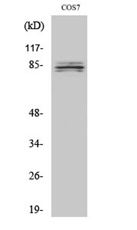
Western Blot analysis of various cells using SEMA4A Polyclonal Antibody
-
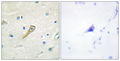
Immunohistochemistry analysis of paraffin-embedded human brain tissue, using SEMA4A Antibody. The picture on the right is blocked with the synthesized peptide.
-
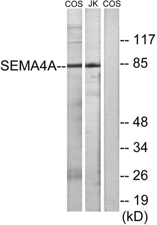
Western blot analysis of lysates from COS7 and Jurkat cells, using SEMA4A Antibody. The lane on the right is blocked with the synthesized peptide.
关闭
在线咨询
Online consultation
-
 在线咨询
在线咨询
-
 技术支持
技术支持

关注微信公众号


关闭